- Electromyography is the technique for evaluating and recording the action potential of muscles. EMG is taken using an instrument called electromyography and the record obtained is known as electromyogram.
- Mainly there are two kinds of EMG measurements .The first method is using surface electrodes and the second one is using needle electrodes .
- In needle or intramuscular EMG is procedure a needle electrode is inserted through the skin into the muscle tissue. A trained expert can observe the electrical activity of muscles when the needle is inserted.
- So in order to obtain the general activity of the muscle cells we use surface electrodes which need to be placed only on the concerned area. So no insertion is required and it will be more comfortable to the patient than needle EMG.
- The electrode used for EMG recording can be of surface type or needle type depending on the area from which the EMG is to be obtained and the type of measurement.
- The bioelectric signals measured from various muscle fibers and neurons are having amplitude ranging from millivolts to microvolts. So it is necessary to amplify the extremely low amplitude EMG signals for the analysis of the data obtained from electrodes.
- During EMG measurement, abnormal and spontaneous activity may be distinguished by the sudden change in sound and this can be analyzed by the physician. The abnormal activity usually indicates muscle damage and they can easily find out the nerve or muscle damage.
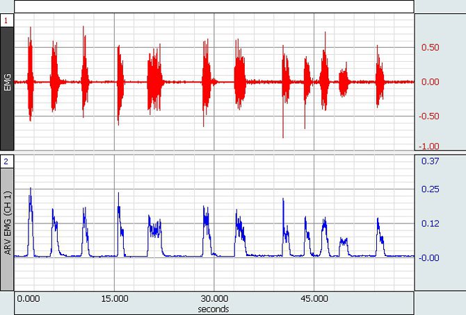
- The measured EMG can be connected to the oscilloscope to visualize the EMG. The abnormalities in the working of nerves and muscle cells can be identified by a physician by analyzing the EMG waveform.
- Due to the low frequency limitation, EMG cannot be recorded on a strip chart recorder because it cannot give a clear idea of the waveform.
- Infected tissues are repaired fastly in diathermy.
- Actually during EMG we are evaluating this bioelectric potential from different cell. This potential is collectively called motor unit action potential (MUAP).
- The shape of the electromyogram is affected by factors such as number of muscle fibers under consideration, the metabolic type of muscle fibers etc.
Author Bio: The Author of this article, Sreejith is writing articles onElectromyogram and Electronics and Communications